
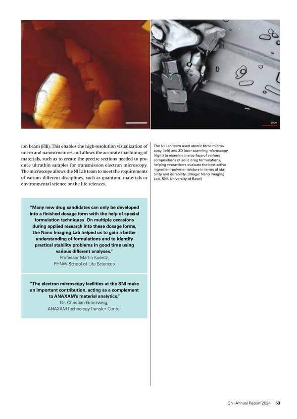
ion beam (FIB). This enables the high-resolution visualization of The NI Lab team used atomic force micros- micro and nanostructures and allows the accurate machining of copy (left) and 3D laser scanning microscopy materials, such as to create the precise sections needed to pro- (right) to examine the surface of various compositions of solid drug formulations, duce ultrathin samples for transmission electron microscopy. helping researchers evaluate the best active The microscope allows the NI Lab team to meet the requirements ingredient-polymer mixture in terms of sta- of various different disciplines, such as quantum, materials or bility and durability. (Image: Nano Imaging Lab, SNI, University of Basel) environmental science or the life sciences. “Many new drug candidates can only be developed into a finished dosage form with the help of special formulation techniques. On multiple occasions during applied research into these dosage forms, the Nano Imaging Lab helped us to gain a better understanding of formulations and to identify practical stability problems in good time using various different analyses.” Professor Martin Kuentz, FHNW School of Life Sciences “The electron microscopy facilities at the SNI make an important contribution, acting as a complement to ANAXAM’s material analytics.” Dr. Christian Grünzweig, ANAXAM Technology Transfer Center SNI Annual Report 2024 53
 Annual Report 2024 - Swiss Nanoscience Institute Page 52 Page 54