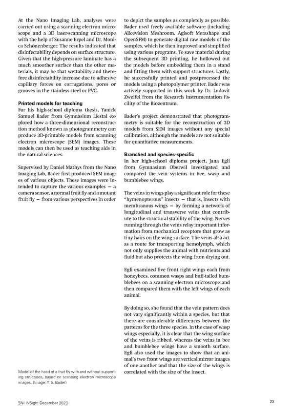
At the Nano Imaging Lab, analyses were to depict the samples as completely as possible. carried out using a scanning electron micro- Bader used freely available software (including scope and a 3D laser-scanning microscope Alicevision Meshroom, Agisoft Metashape and with the help of Susanne Erpel and Dr. Moni- OpenSFM) to generate digital raw models of the ca Schönenberger. The results indicated that samples, which he then improved and simpli昀椀ed disinfectability depends on surface structure. using various programs. To save material during Given that the high-pressure laminate has a the subsequent 3D printing, he hollowed out much smoother surface than the other ma- the models before embedding them in a stand terials, it may be that wettability and there- and 昀椀tting them with support structures. Lastly, fore disinfectability increase due to adhesive he successfully printed and postprocessed the capillary forces on corrugations, pores or models using a photopolymer printer. Bader was grooves in the stainless steel or PVC. actively supported in this work by Dr. Ludovit Zweifel from the Research Instrumentation Fa- Printed models for teaching cility of the Biozentrum. For his high-school diploma thesis, Yanick Samuel Bader from Gymnasium Liestal ex- Bader’s project demonstrated that photogram- plored how a three-dimensional reconstruc- metry is suitable for the reconstruction of 3D tion method known as photogrammetry can models from SEM images without any special produce 3D-printable models from scanning calibration, although the models are not suitable electron microscope (SEM) images. These for quantitative measurements. models can then be used as teaching aids in the natural sciences. Branched and species-specific In her high-school diploma project, Jana Egli Supervised by Daniel Mathys from the Nano from Gymnasium Oberwil investigated and Imaging Lab, Bader 昀椀rst produced SEM imag- compared the vein systems in bee, wasp and es of various objects. These images were in- bumblebee wings. tended to capture the various examples — a camera sensor, a normal fruit 昀氀y and a mutant The veins in wings play a signi昀椀cant role for these fruit 昀氀y — from various perspectives in order “hymenopterous” insects — that is, insects with membranous wings — by forming a network of longitudinal and transverse veins that contrib- ute to the structural stability of the wing. Nerves running through the veins relay important infor- mation from mechanical receptors that grow as tiny hairs on the wing surface. The veins also act as a route for transporting hemolymph, which not only supplies the animal with nutrients and 昀氀uid but also protects the wing from drying out. Egli examined 昀椀ve front right wings each from honeybees, common wasps and bu昀昀-tailed bum- blebees on a scanning electron microscope and then compared them with the left wings of each animal. By doing so, she found that the vein pattern does not vary signi昀椀cantly within a species, but that there are considerable di昀昀erences between the patterns for the three species. In the case of wasp wings especially, it is clear that the wing surface of the veins is ribbed, whereas the veins in bee and bumblebee wings have a smooth surface. Egli also used the images to show that an ani- mal’s two front wings are vertical mirror images of one another and that the size of the wings is Model of the head of a fruit 昀氀y with and without support- correlated with the size of the insect. ing structures, based on scanning electron microscope images. (Image: Y. S. Bader) SNI INSight December 2023 23
 SNI INSight December 2023 Page 22 Page 24