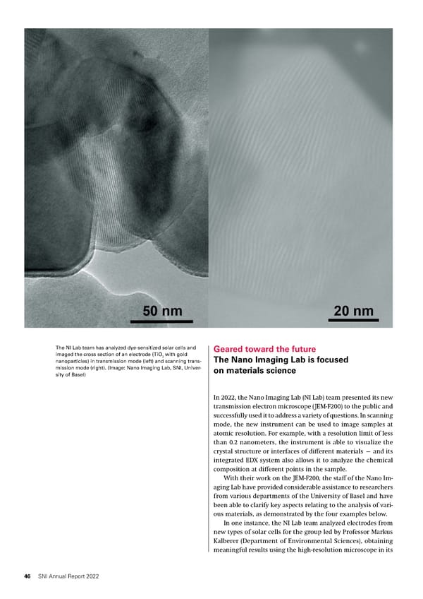
The NI Lab team has analyzed dye-sensitized solar cells and Geared toward the future imaged the cross section of an electrode (TiO with gold 2 The Nano Imaging Lab is focused nanoparticles) in transmission mode (left) and scanning trans- mission mode (right). (Image: Nano Imaging Lab, SNI, Univer- on materials science sity of Basel) In 2022, the Nano Imaging Lab (NI Lab) team presented its new transmission electron microscope (JEM-F200) to the public and successfully used it to address a variety of questions. In scanning mode, the new instrument can be used to image samples at atomic resolution. For example, with a resolution limit of less than 0.2 nanometers, the instrument is able to visualize the crystal structure or interfaces of different materials — and its integrated EDX system also allows it to analyze the chemical composition at different points in the sample. With their work on the JEM-F200, the staff of the Nano Im- aging Lab have provided considerable assistance to researchers from various departments of the University of Basel and have been able to clarify key aspects relating to the analysis of vari- ous materials, as demonstrated by the four examples below. In one instance, the NI Lab team analyzed electrodes from new types of solar cells for the group led by Professor Markus Kalberer (Department of Environmental Sciences), obtaining meaningful results using the high-resolution microscope in its 46 SNI Annual Report 2022
 SNI Annual Report 2022 Page 45 Page 47