
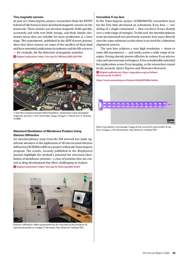
Innovative X-ray lens In the Nano-Argovia project ACHROMATIX, researchers have for the first time developed an achromatic X-ray lens — con- sisting of a single component — that can focus X-rays sharply over a wide range of energies. To this end, the interdisciplinary team incorporated two previously separate lens types directly onto the same substrate so that there is no need for a laborious alignment process. The new lens achieves a very high resolution — down to some 200 nanometers — and works across a wide range of en- ergies. Having already proven effective in various X-ray micros- copy and spectroscopy techniques, it has considerable potential for applications across X-ray imaging, as the researchers report in the journals Optics Express and Photonics Research. Original publications: https://opg.optica.org/oe/fulltext. cfm?uri=oe-33-12-26578 https://www.researching.cn/Articles/OJb3d672085c7e2a4e Tiny magnetic sensors As part of a Nano-Argovia project, researchers from the FHNW School of Life Sciences have developed magnetic sensors on the nanoscale. These sensors can measure magnetic fields quickly, accurately and with very little energy, and their simple elec- tronics mean they are suitable for mass production at a later stage. The experiments, published in the IEEE Sensors Journal, show that these sensors are some of the smallest of their kind and have potential applications in industry and the life sciences — for example, for the detection of magnetic particles. Original publication: https://doi.org/10.1109/jsen.2025.3537700 In the Nano-Argovia project NanoHighSens, researchers have developed magnetic sensors in the nanometer range. (Image: J. Pascal and H. Nicolas, FHNW) Structural Elucidation of Membrane Proteins Using Electron Diffraction An interdisciplinary team from the SNI network has made sig- nificant advances in the application of 3D microcrystal electron diffraction (3D ED/MicroED) in a project within the Nano-Argovia program. The results, recently published in the Biophysical Journal, highlight the method’s potential for structural eluci- dation of membrane proteins – a class of proteins that are cen- tral to drug development but often challenging to analyze. Original publication: https://doi.org/10.1016/j.bpj.2025.10.027 Electron diffraction offers great potential for elucidating the structure of membrane proteins. (Image: V. Panneels, Paul Scherrer Institute PSI) Scanning electron microscope image of the monolithic achromatic X-ray lens. (Image: J. Vila-Comamala, Paul Scherrer Institute PSI) 35 SNI Annual Report 2025
 Annual Report 2025 - Swiss Nanoscience Institute Page 34 Page 36